 Contact Us
We analyze brain/neural data by harnessing the power of AI technologies.
Brain imaging like MRI and EEG has enormous potential for diagnosing and predicting brain disease and mental illness. However, the data obtained from just one person’s brain is large and complex and requires a huge amount of effort to prepare a proper environment and conduct cutting-edge analyses.
Thus, ARAYA provides setup services for automatic analysis environments and analysis services using AI/machine learning technologies to research institutes. Our services enable you to conduct cutting-edge analyses just by pushing a button, without worrying about complicated programming or tedious computer setup. In addition, we also help our customers to construct AI/machine learning models that predict individual characteristics, symptoms, and health indexes from brain data.
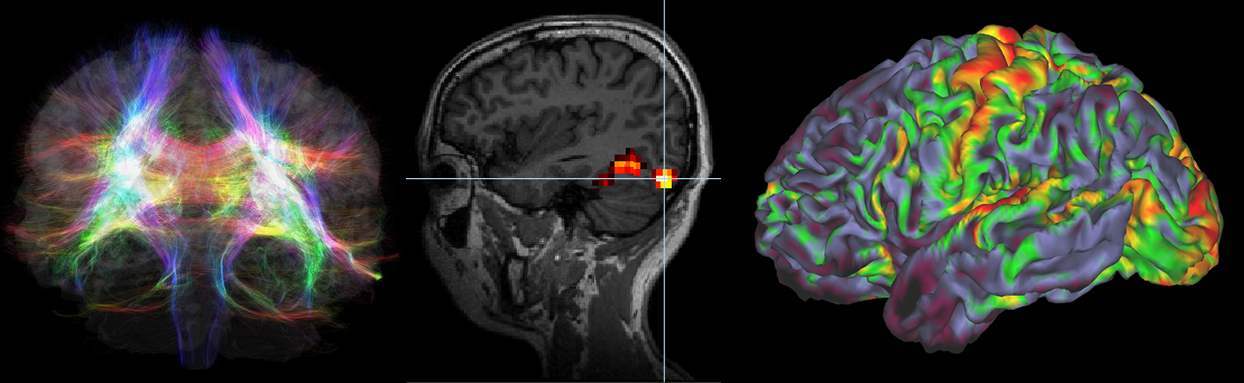
1. Automatic analysis of MRI data
2. Constructing predictive models using brain data
3. Assist in utilizing brain characteristics data for product development
4. Neuromarketing
5. Developing an effective teaching method (e.g., language learning)
6. Consultation in neuroscience research planning
-Structural MRI
-Diffusion MRI
-Functional MRI (task-fMRI, resting state-fMRI)
-Quantitative MRI
-EEG (Electroencephalogram)
-MEG (Magnetoencephalography)
Case studies
For the following research papers, ARAYA assisted brain data analyses.
Sugawara, A., Katsunuma, R., Terasawa, Y., & Sekiguchi, A. (2024). Interoceptive training impacts the neural circuit of the anterior insula cortex. https://doi.org/10.1038/s41398-024-02933-9
Tose, K., Takamura, T., Isobe, M., Hirano, Y., Sato, Y., Kodama, N., ... Sekiguchi, A. (2024). Systematic reduction of gray matter volume in anorexia nervosa, but relative enlargement with clinical symptoms in the prefrontal and posterior Molecular Psychiatry. https://doi.org/10.1038/s41380-023-02378-4
Orihashi, R., Mizoguchi, Y., Imamura, Y., Yamada, S., Ueno, T., & Monji, A. (2020). Oxytocin and elderly MRI-based hippocampus and amygdala volume: A 7-year follow-up study. Brain Communications, 2 (2). Brain Communications, 2(2). https://doi.org/10.1093/braincomms/fcaa081
Sugawara, A., Terasawa, Y., Katsunuma, R., & Sekiguchi, A. (2020). Effects of interoceptive training on decision making, anxiety, and somatic symptoms. https://doi.org/10.1186/s13030-020-00179-7
Hagiwara, A., Hori, M., Kamagata, K., Warntjes, M., Matsuyoshi, D., Nakazawa, M., ... Aoki, S. (2018). Myelin measurement: comparison between simultaneous tissue relaxometry, magnetization transfer saturation index, and t1w/t2w ratio methods. Scientific Reports, 8 (1). Available at: https://doi.org/10.1038/s41598-018-28852-6
Hagiwara, A., Hori, M., Yokoyama, K., Nakazawa, M., Ueda, R., Horita, M., ... Aoki, S. (2017). Analysis of white matter damage in patients with multiple sclerosis via a novel in vivo MR method for measuring myelin, axons, and g-ratio. American Journal of Neuroradiology, 38 (10), 1934-1940. https://doi.org/10.3174/ajnr.a5312
